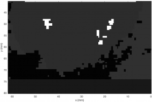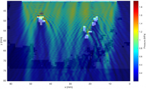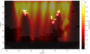
When ultrasound propagates in the human body, it travels through several layers of different tissues which affect the energy and direction of the waves. The combined effect of all these layers results in the loss of intensity in the therapeutic ultrasound field due to reflection, refraction, dispersion and attenuation of acoustic energy. This reduction in ultrasound intensity in turn decreases the heating efficacy of the ultrasound field, which may result in untreated areas in the target tumour.
Different tissue layers and abnormalities can significantly reduce the efficiency of the ultrasound therapy in the target organ. For a patient, this would mean either longer therapy times, or in the worst case, the whole treatment would have to be terminated and conducted again. Either case would mean inconveniences for both the patient and clinical personnel. The aim of using computational models to simulate ultrasound therapy is to predict the treatment outcome in different scenarios in order to avoid these situations.
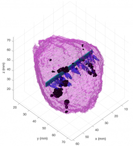
Whilst it is difficult (if not impossible) to measure all the effects in vivo in the tissue, it is possible to replicate all the scenarios using three-dimensional clinical image data from patients together with suitable computational models. Hence, the research is carried out using accurate acoustic and thermal simulation models together with the patient tomography data in order to realistically predict the treatment outcome. The patient data used in the computational simulations are acquired during the actual ultrasound treatment at the hospital and saved for further analysis. The results from the real treatment and the computational model are then compared to draw conclusions and suggest improvements to the current ultrasound therapy protocol.
The big benefit of conducting the simulations with clinical patient data at the hospital is that the results can be immediately applied to the planning stage of the next treatment. The results can then be used to predict the treatment outcomes before the actual therapy as well as determine criteria whether the patient would benefit from the ultrasound therapy or whether alternative treatment options should be considered.