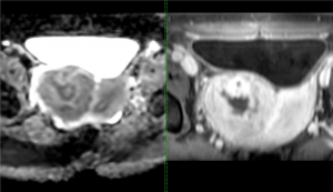
Therapeutic ultrasound treatment result depends on tissue properties e.g. density and the intensity attenuation coefficient. The suitability of the tumor is usually evaluated beforehand qualitatively from MR images. Qualitative evaluation is not reliable because the signal of the MR images can vary greatly due to imaging parameters which increases the risk of not achieving the optimal treatment result.
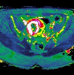
The suitability could be evaluated more reliably with quantitative MRI techniques because in quantitative MRI the scanner is not only used to scan but also to measure quantitative parameters from the tissues. In that case the 3-D pixels (voxels) in the MR images consist of unambiguous values instead of variable signal intensity. These kind of tissue parameters are T1- and T2-relaxation times, proton density, apparent diffusion coefficient, pseudo diffusion coefficient and perfusion values. In previous studies they have noticed that some of the quantitative MRI techniques could be used to evaluate the suitability of uterine fibroids and bone metastases.
The possible benefits of using the quantitative MRI techniques in clinical practice are better treatment outcomes, not having any inconveniences for both the patient and clinical personnel and treatment would be more cost-effective. The aim of this research is to develop MRI based evaluation method to predict the outcome of therapeutic ultrasound treatments.